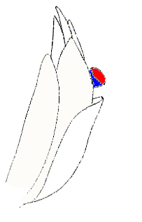
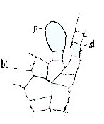
Case StudiesHymenoscyphus schimperiThe fungus Hymenoscyphus schimperi is an apothecial ascomycete that parasitizes the northern hemisphere moss Sphagnum squarrosum. If you are looking for more information about this fungus look for it also under the names Helotium schimperi and Discinella schimperi. The images on this page come from the 1888 paper by Nawaschin that is given in the references at the end of this page. I have added a few colours and arrows to better highlight some features. The apothecia are under a millimetre in diameter and the left-hand diagram below, of part of a Sphagnum plant, shows a cluster of leaves with one coloured apothecium of Hymenoscyphus schimperi visible. Though the apothecia may develop on the leaves the fungus does not infect the leaves. In the axils between the leaves and stems are mucilage-producing papillae and it is these papillae that the fungus infects. On the left of the right-hand diagram (labelled with the letters bl) is the base of a Sphagnum leaf and on the right (labelled with the letters st) is part of the Sphagnum stem from which the leaf has grown. Between the stem and the leaf is the papilla, labelled with the letter p.
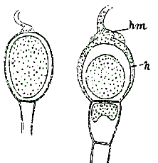
The infection begins when a single hypha makes contact with the papilla. The fungus then produces a pad-like appressorium of much-branched, closely-packed hyphae over the upper part of the papilla. At the same time the fungus also penetrates the surface of the papilla and a much-branched haustorium is formed inside the papilla cell wall. The following pair of diagrams shows two papillae in cross-section. The papilla on the left is shown just before fungal infection. You can see a fungal hypha that has just reached the top of the papilla. On the right is fungally-infected papilla. You can see the pad-like appressorium (labelled with the letters hm) on the outer surface of the papilla's apex. Additionally the fungal hyphae have penetrated the papilla and formed a hyphal mass (labelled h) between the outer and inner walls of the papilla.
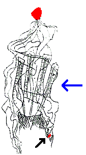
In the next diagram the tiny red dot (near the tip of the black arrow at the bottom of the diagram) shows an infected papilla. The numerous wavy lines indicate some of the fungal hyphae that have grown out over the Sphagnum plant to produce an apothecium (the upper red patch). In order to produce this drawing Nawaschin carefully removed the surrounding leaves, taking care to limit any damage to the hyphal network. Once the hyphal network had been exposed Nawaschin was able to observe the hyphae that connected the fungal fruiting body and the Sphagnum papilla. The blue arrow points to four Sphagnum archegonia.
|
![An Australian Government Initiative [logo]](/images/austgovt_brown_90px.gif)


