|
Ecology
Foliicolous lichens
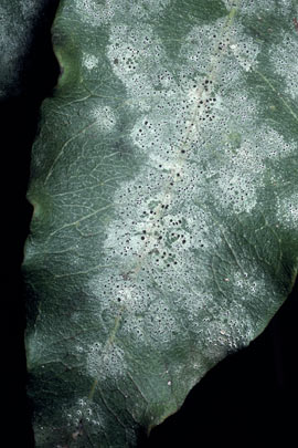
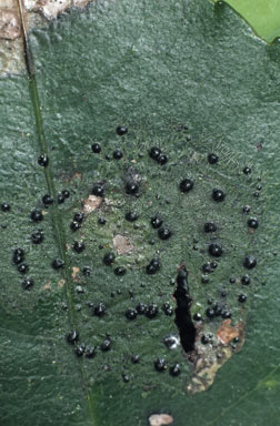
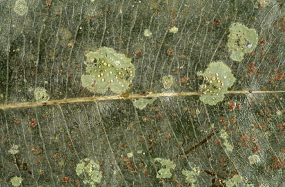
Foliicolous (or epiphyllous) lichens are those that grow on the leaves of vascular plants. Such lichens are widespread and are especially common in the tropical areas where there are long periods of high humidity. Many foliicolous species are strictly foliicolous but some may also be found on other plant parts (twigs, branches, trunks) or even non-plant substrates such as rocks. Apart from lichens you can also find algae, cyanobacteria, bryophytes and fungi growing on vascular plant leaves. The living space on the leaf surface is referred to as the phyllosphere and from what's already been said about the variety of epiphylls (a general term for organisms living on leaves) it's clear that the phyllosphere can be a complex micro-habitat. Much is still unknown about the roles and interactions of foliicolous lichens.
At first it would seem that anything growing on a plant leaf must be detrimental to that plant and the following are some of the theoretical arguments that have been advanced to support that hypothesis. If part of a leaf is covered the total leaf photosynthesis is reduced so that there is less food production. The rhizines that anchor a lichen to its host leaf, may penetrate the leaf and so provide potential entry points for pathogenic organisms. Moreover, moisture held by the lichens creates a more humid microhabitat on the leaf surface, potentially ideal for various pathogenic micro-organisms. As plausible as such arguments may sound they must still be tested, rather than automatically accepted, and the reality is not necessarily what the arguments suggest. A case in point is the argument about epiphyll cover reducing leaf photosynthesis. In fact there may not be much effect on plant photosynthesis. This was the subject of a study in Queensland's Mossman Gorge National Park that looked closely at the effects of lichens on leaves of Calamus australis, a palm, and Lindsayomyrtus racemoides, a plant in the family Myrtaceae. The latter species was formerly known by the name Eugenia racemoides. Eleven lichen species were involved and the amount of light transmitted through the lichens to the underlying leaf varied from about 30 to 70 percent of the incident light, depending on the lichen species. The researchers found that the host leaves compensated for the lichens and that total photosynthesis was not reduced. In both host plants the chlorophyll content in those parts of the leaves colonized by lichens was significantly higher than in uncolonized leaf areas. Of course, these results need not hold for other species of lichens and perhaps not for other vascular host species. However the lichen study confirms that the simplistic theoretical conclusion about photosynthesis, given early in this paragraph, cannot hold in general![]() .
.
Thus far you could be excused for concluding that, while host leaves can at best tolerate epiphylls, there's not really anything in it for the hosts. In another study undertaken in Costa Rica two researchers carefully removed liverworts and lichens from randomly selected leaves of grapefruit (Citrus paradisi) and an understorey cyclanth (Cyclanthus bipartitus). They found that the denuded leaves suffered 2 to 3 times more damage by leafcutter ants (“one of the most destructive herbivores in neotropical rainforests”) than was the case with the covered leaves. Presumably the chemicals produced by the liverworts or lichens are harmful either directly to the ants or to the ants’ fungus gardens. Liverworts and lichens both produce a considerable array of chemical compounds but the study did not try to differentiate lichen from liverwort effects![]() .
.
Leaves do not form a static habitat. They emerge, grow in size and after some time fall off. As a plant grows in height or bulk leaves that were once exposed to light can become shaded. Water running off higher leaves may carry leached nutrients to the lower leaves. These and other factors can work to create a variety of changing MICROHABITATS. Wilkiea macrophylla is found in rainforest areas from northern Queensland south to New South Wales. In the rainforests of south-eastern Queensland the species is common as a shrub or small tree in the understorey and the leaves, which are about ten centimetres long and four centimetres wide, with a mean half-life estimated at 6.8 years. In one study leaves of various ages were collected and their lichen cover analyzed. Aderkomyces albostrigosus and Sporopodium xantholeucum appeared to be pioneer lichen species on young leaves. Though other species also could begin colonization of young leaves at the same time, the evidence suggested that those two matured more quickly and showed best thallus development when thalli of other species were not growing close by. Older leaves showed good growth by Porina epiphylla, Porina impressa and Mazosia melanophthalma. Separate thalli of Porina epiphylla could fuse to create larger, composite thalli - and similarly with Porina impressa. On well-colonised leaves Aderkomyces albostrigosus and Sporopodium xantholeucum were less frequent and their thalli appeared less healthy. Thalli of Mazosia melanophthalma grew as discrete, round patches but showed no signs of necrosis when surrounded by Porina. This Mazosia species therefore appears better able to withstand competition than do Aderkomyces albostrigosus and Sporopodium xantholeucum. Strigula orbicularis and Strigula subtilissima establish at leaf wounds. Since the probability of leaf damage increases with leaf age it's not surprising that the Strigula species appeared with greater frequency on older leaves. Lichen cover varied greatly from leaf to leaf and between taxa. Coverage could reach 100% of the leaf surface, though rarely. Generally coverage increased with leaf age and the older leaves had a mean coverage of about 50%. The greatest observed percentage coverage by a single species on any one leaf was an instance where Porina epiphylla covered 78% of one leaf surface, but this was an extreme. For example, on the older leaves mean coverage by this species was about 30%. At the other extreme there were a number of species where each had a maximum coverage of under 10% of the leaf surface. Incidentally, the studies of Wilkiea macrophylla showed that lichens made a distinction between the two leaf surfaces. Lichens were found only on the morphological upper surface of the leaf even if, by some accident, a leaf had been inverted to leave the morphological lower surface facing upward![]() .
.
Foliicolous lichens and studies of early thallus development
The processes of propagule germination and thallus development have been studied in relatively few lichens and then mostly under laboratory, rather than natural, conditions. In theory foliicolous lichens should be good candidates for studies of thallus development since they exploit relatively short-lived substrates. Hence the thalli must be able to develop fairly rapidly but when the thalli grow attached to leaves it is hard to detect and examine the early stages of development. It has been found that many foliicolous lichens will grow on various artificial substrates and two researchers investigated in situ propagule germination and thallus growth at a site in a lowland Brazilian forest, via the following method:
Plastic screening with a mesh of about 2 mm square was cut into strips 3-4 cm wide and 20-35 cm long. Plastic microscope cover slips (20 x 20 mm) were fastened to the strips of screening by inserting their corners into small diagonal slits cut into the mesh...The strips were tied, with the cover slips facing upward, to the upper surfaces of leaves of Bactris sp., a common understorey palm which typically bears well-developed foliicolous lichen communities... At intervals of 3-6 wk, some of the cover slips were removed for microscopic examination. Debris and microorganisms were wiped off the lower surface of the cover slips, which were then inverted and mounted with distilled water onto clean microscope slides. In a few instances, a clearer image was obtained by placing the colonized cover slip face up on a microscope slide, with a glass cover slip mounted over it
.
The use of a clear plastic substrate eliminated the problem of distinguishing lichen from leaf tissue and also made it easier to see germinating spores or the very early stages of thallus development. The researchers did try to make sequential observations of the same individuals, by returning the cover slips to the field but these attempts failed. The intense light of the microscope lamp may have damaged the photobionts. Within 3-5 weeks after the start of the experiment algal cells and germinating lichen propagules were visible on many cover slips. After 6 months the cover slips had developed such a covering of lichens, other organisms and debris that clear microscopic observation was no longer possible.
The cover slips were host to a variety of propagules - fungal spores (with or without attached photobiont cells), lichenized vegetative propagules (such as soredia or isidia) or fungal vegetative propagules. Conidia are the best known fungal vegetative propagules. They are small, easily dispersed propagules, produced by various processes (e.g. budding or hyphal fragmentation) and often in small chambers called pycnidia. Various lichenized fungi in the family Gomphiliaceae produce propagules called diahyphae which consist of wefts of hyphae or short chains of fungal cells. Diahyphae are also examples of vegetative fungal propagules and propagules that are much more substantial than conidia.
Developing thalli showed different methods of mycobiont-photobiont co-ordination. In a genus such as Tricharia the algal partner has spherical cells and the
...phycobiont [i.e. algal] cells were organized at the periphery of the young lobes, their spherical cells positioned at the tips of parallel, longitudinally appressed hyphae growing outward from the thallus. This type of organization appears to provide a very simple mechanism by which the growth of the mycobiont directly distributes the dividing phycobiont cells to the youngest parts of the radially expanding thallus. Inasmuch as the algal cells and their division products remain positioned at the tips of the advancing hyphae and their branches, apical growth of the hyphae would be sufficient to move the algal cells...
By contrast the photobiont of Coenogonium is filamentous. The algal filaments give the impression of autonomous growth and branching, but algal growth followed radial hyphal growth fairly strictly. The researchers suggested that algal growth is controlled by substances released from the hyphae.
The researchers were able to observe interactions between the mycobionts of neighbouring thalli. It had long been known that mycelia of non-lichenized fungi can interact in various ways when they meet. One mycelium may inhibit further growth of the other and thereby occupy more area, one may damage or destroy part or all of the other (sometimes by chemical means) or there may be a stand-off, with neither able to grow further into the other's territory. In the last case each mycelium would then put effort into those areas where further growth was possible and, in effect, "seal off" the area in contact with the other mycelium. There was no reason to suppose that lichenized fungi would not display a variety of interactions. Where several lichens grow together it is often easy to see that the thallus of one species will overgrow the thallus of another, showing clearly that one is a more aggressive competitor. However, previously there had been no microscopic studies in this area but the ease of viewing through a transparent substrate allowed the researchers to produce evidence of the inhibitory effects of one mycobiont on another.
More information
Robert Lücking's monograph Foliicolous lichenized fungi, published in 2008 by the New York Botanical Garden as volume 103 in the Flora Neotropica series, is ostensibly a taxonomic study of the foliicolous lichens of the American tropics. Certainly the bulk of the volume's 866 pages is devoted to species descriptions. However, the first 100 pages give much information about the structures, classification and ecology of the foliicolous lichens and would be useful to anyone interested in foliicolous lichens.
You will find a hundred photographs of foliicolous lichens here.
 |
(left) Byssoloma sp. (above) Sporopodium vezdeanum (right) Strigula smaragdula |
 |
![An Australian Government Initiative [logo]](/images/austgovt_brown_90px.gif)


